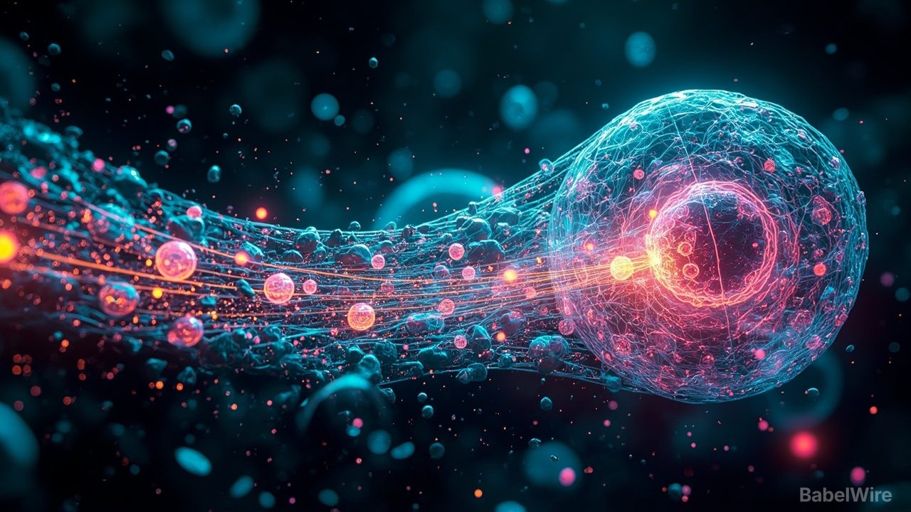
Quanta Magazine has published a set of images showing the inside of cells with a clarity that would have looked like illustration a generation ago. The immediate reaction to work like this is aesthetic — the pictures are genuinely beautiful, color-coded and luminous, and they circulate far beyond the labs that made them. The more interesting reaction is structural: images like these are how a field discovers that its questions have changed.
It is worth remembering how hard the underlying problem is. A cell is mostly water and protein, and water and protein bend light almost identically, which means a living cell is nearly transparent. For most of the history of microscopy, seeing inside one meant killing it, fixing it, slicing it thin, and staining it — a procedure that produced exquisite detail and destroyed the thing you wanted to understand. The structure survived; the dynamics did not.
The advances that changed this fall into a few families, and the current gallery is best read as a demonstration of what they make visible together:
Beating the diffraction limit. For over a century, the resolution of a light microscope was assumed to be capped at roughly half the wavelength of the light used — a barrier that made many cellular structures permanently unresolvable, no matter how good the lens. Super-resolution methods broke that assumption by treating fluorescence as something to be controlled over time rather than simply collected: switching individual molecules on and off, then computing the position of each, turns a blur into a map. Structures that textbooks once drew as schematics can now be measured.
Imaging without fixing. Cryogenic methods freeze a cell fast enough to preserve its molecular architecture and then image it in three dimensions at near-atomic detail, capturing the cell as it actually was at the instant of freezing rather than as a stained approximation. Cryo-electron tomography has become the standard way to see the crowded interior of a cell the way it really is — not the tidy diagrams, but a dense, jostling environment where everything touches everything else.
Expanding the sample instead of the optics. A counterintuitive trick: rather than improving the microscope, physically swell the tissue by embedding it in a polymer that expands, spreading molecules apart until ordinary optics can resolve them. Expansion microscopy converted a hardware problem into a chemistry problem, which made high-resolution imaging available to labs that could never afford the hardware route.
Watching it happen. The most consequential shift is temporal. Fluorescent proteins and gentler illumination let researchers follow a process — a vesicle moving, a protein assembling, a signal propagating — over minutes or hours in a living cell. Structural biology's classic output is a shape; live imaging's output is a mechanism, and mechanisms are what drug discovery actually needs to intervene in.
Why the pictures matter beyond publicity: they are how a field's capabilities become legible to people outside it. A researcher deciding which technique to invest in, a funder deciding what to fund, a student deciding what to study — all of them calibrate on what they have seen is now possible. Vivid, well-chosen images do real work in that calibration, which is why serious journals and magazines keep commissioning them rather than treating them as decoration.
There is a fair caution. Beautiful images can imply a clarity that the underlying data does not always have: color is almost always added, structures are segmented by software whose assumptions matter, and a single frame of a dynamic process can mislead about what is typical. The honest reading of any such gallery is that it shows what the best current instruments can capture in skilled hands — a ceiling of possibility, not an average of what every experiment sees.
Still, the direction is unambiguous. Biology spent decades describing the parts list of the cell and is now in the business of watching the parts work. Every improvement in resolution, speed, and gentleness moves the field toward the thing it has always wanted: a cell observed while it is still alive, still crowded, and still doing whatever it does when nobody has fixed it.
What to watch: how fast cryo-tomography workflows become routine rather than heroic, whether live-cell imaging extends to whole organisms at useful resolution, and whether the analysis software — the segmentation and reconstruction that turns pixels into claims — keeps pace with the instruments that produce them. The last one is the quiet bottleneck, and the least photogenic.