For decades, sleep research treated the cerebral cortex as a passive receiver — the place where deep-sleep brain waves are visible, but not where they originate. A new Nature study upends that assumption by identifying a tiny population of cortical cells that can actively trigger sleep.
The cells, called Sst-Chodl neurons, make up roughly 0.1% of all cortical neurons — about one in a thousand. They are defined by the co-activity of two genes (Sst and Chodl), and targeting them required years of work because studying either gene alone captures a far larger, heterogeneous population of neurons.
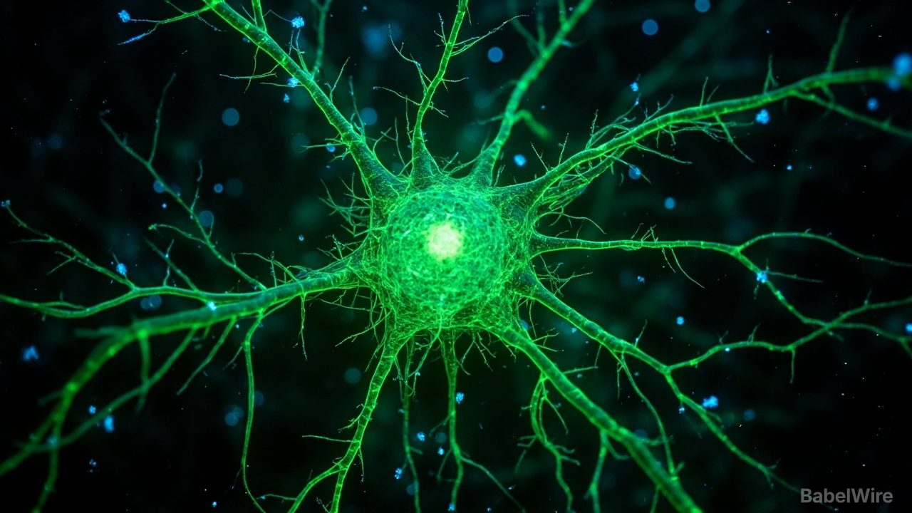
Renata Batista-Brito, who runs the lab at Albert Einstein College of Medicine, described the anatomy as immediately striking despite the rarity. "At first, I saw two or three cell bodies in the whole brain," she told Ars Technica. "Despite that, there was massive, massive arborization all over the visual cortex, like I never saw with any other neuron."
That branching pattern is the opposite of typical inhibitory neurons, which receive broad inputs but act locally. Sst-Chodl neurons receive precise inputs but broadcast widely — a single cell can branch across the entire visual cortex and send axons to areas governing touch, hearing, spatial memory, navigation, and voluntary movement.
When the team imaged these cells while tracking mouse behavior, 95 out of 111 cells fired during slow-wave sleep and quiet wakefulness, going silent during running and REM sleep. During slow-wave sleep, the cells peaked precisely when other neurons wound down — at the termination of UP states — suggesting a coordinating role.
Using optogenetics to activate the cells on demand, the researchers drove delta power — the slow, high-amplitude brain waves of deep sleep — up across every cortical layer. Crucially, the frequency was intrinsic: flat stimulation, delta-frequency, 20 Hz, or 60 Hz all produced the same oscillation pattern. "There's something about the intrinsic properties of these cells that, once they trigger, they go on this one frequency," Batista-Brito said.
The behavioral results were dramatic. Activating Sst-Chodl neurons across the cortex increased slow-wave and REM sleep, reduced time to fall asleep, and sent mice to their nests during the daytime. Even during the dark phase — when nocturnal mice are normally active — the team could make them sleep more than they usually do during the day.
The cells are conserved from salamanders to humans, which the authors argue makes them a potential entry point for understanding sleep disruption in psychiatric illness. Three open questions remain: what normally activates these cells, whether they truly sense homeostatic sleep pressure, and why they specifically drive delta oscillations.