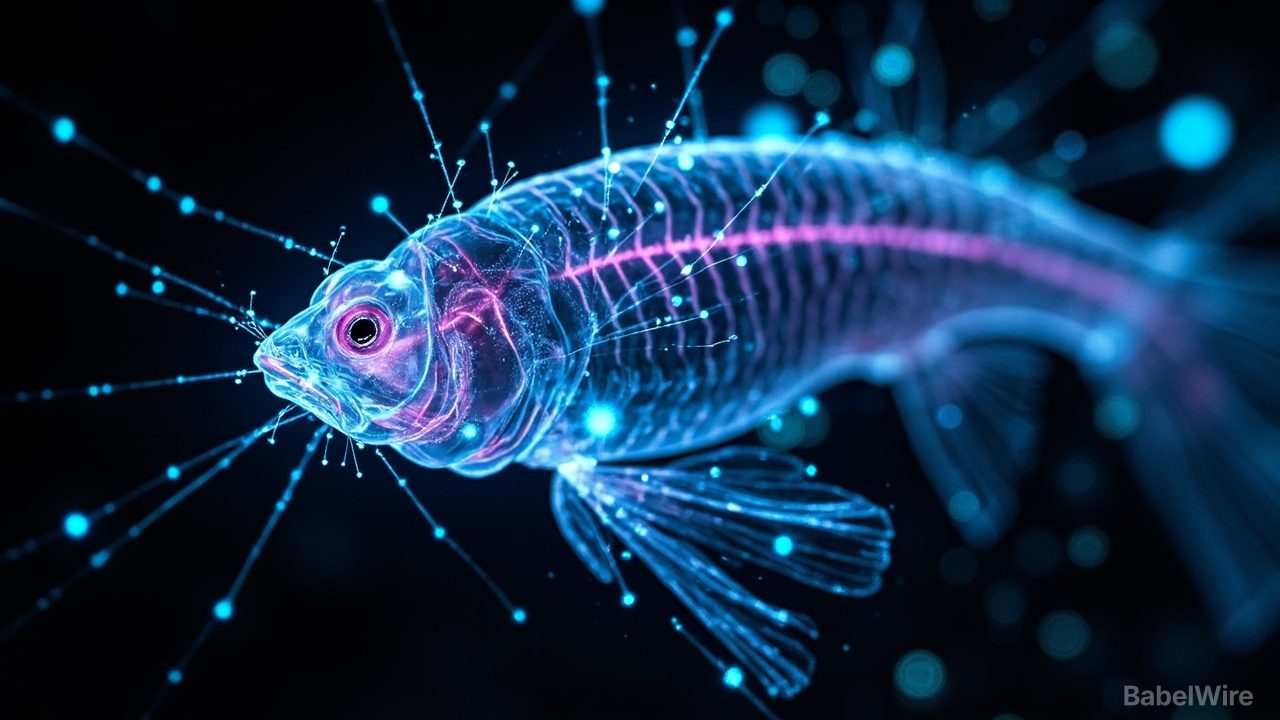
MIT engineers have built a high-speed microscope that can image electrical activity in neurons across the entire brain of a living animal — capturing 200 complete scans per second of a larval zebrafish's whole brain. The technique, unveiled Aug. 14, could help scientists learn how the entire brain works together to generate decisions and emotions.
Neurons communicate with electrical signals, but watching that activity in real time across large brain regions has been a major challenge. Conventional microscopes are too slow to catch the rapid, tiny electrical flickers that ripple through neural circuits. The new instrument solves the problem with a modified light-sheet microscope that combines ultrashort pulses of laser light with a highly sensitive detector to sweep across the larva's entire head.
Because electrical signals in neurons can be coupled to light emission — through fluorescent proteins that respond to voltage changes — the microscope effectively films electrical activity as it happens. At 200 frames per second, it can catch the fast waves of neural activity that drive behavior.
In experiments with larval zebrafish, the researchers recorded activity patterns across the whole brain while the animals moved freely, watching individual neurons fire as the fish chased prey or dodged obstacles. The team also showed the technique is sensitive enough to detect single electrical impulses in individual neurons.
The invention, described in a study released Aug. 14, moves neuroscience closer to a long-held goal: seeing how the whole brain operates as one integrated system. Because zebrafish share many fundamental brain circuits with mammals, the insights may one day extend to more complex brains — helping to decode how decisions, learning and emotion emerge from billions of neurons.