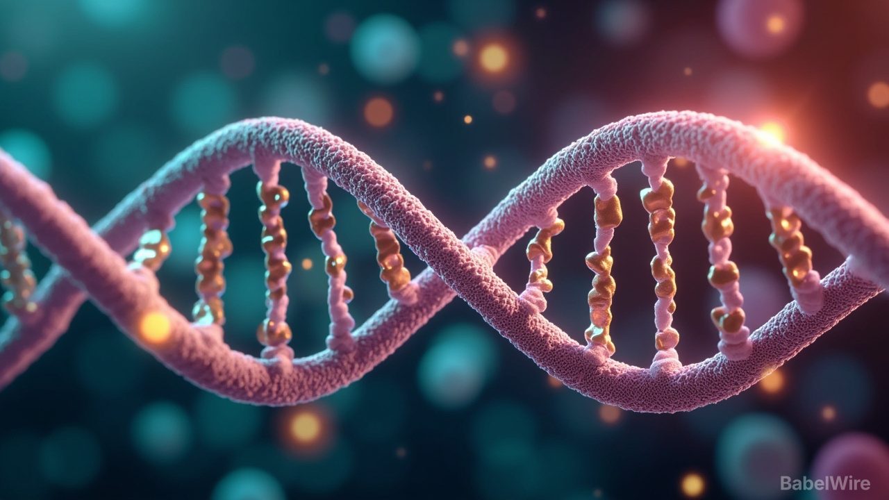
Physicists have captured something biologists have argued about for two decades: two DNA double helices zipping together, groove for groove, and the invisible scaffolding that allows it to happen.
DNA molecules carry the same negative charge, so they should push each other apart. They nevertheless pair up inside cells, a step that matters for genetic recombination, gene silencing and the emergence of cancer. Researchers at the University of York and the University of Sheffield used high-powered atomic force microscopy to watch short DNA fragments align, then ran atom-level computer simulations to work out why.
The answer, published in Nucleic Acids Research, is divalent metal ions. The simulations show that ions carrying two positive charges sit inside the grooves of the helices and act as tiny bridges, holding both strands at once across the gap. The images confirm a model known as the DNA zipper, proposed around twenty years ago by Alexey Kornyshev, then at Imperial College London, which predicted that salt ions create alternating charge patterns allowing helices to interlock like spiral staircases.
The work also shows that pairing is not uniform. Some sequences form contacts far stronger than others, creating hotspots where two helices are especially likely to line up — exactly the kind of genomic region that may matter when mutations disrupt normal cellular processes.
"This discovery could help researchers identify regions of the genome specially involved in DNA pairing," said Agnes Noy of York, who co-led the research. "These regions may become particularly important when mutations disrupt normal cellular processes and contribute to cancer."
Beyond cancer biology, the finding points to programmable interactions that engineers could eventually exploit to design custom DNA structures. "It was incredible to be able to directly visualise the long-hypothesised mechanism for the first time," said co-lead author Thomas Catley of Sheffield.