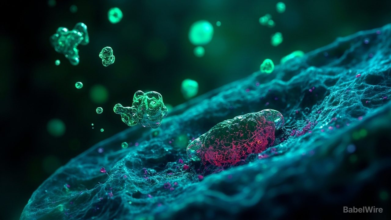
For more than half a century, collagen has been depicted as a long, rigid molecular cable — the structural protein that gives skin, bones, tendons, and organs their strength. But that familiar image may capture only what collagen becomes after it leaves the cell, not how it actually exists while being made.
Using high-resolution imaging of living cells, researchers at the Center for Genomic Regulation (CRG) in Barcelona have now observed collagen in a strikingly different form: as soft, liquid-like droplets rather than stiff rods.
Published in the Journal of Cell Biology, the study provides the first direct view of the most abundant protein in the human body in its natural intracellular state. Collagen makes up roughly one-third of the body's total protein mass.
"Inside a cell, collagens are not rigid molecules as one had assumed. They are in fact, very pliable, taking a liquid condensate form much like oil in a drop of water," explains ICREA Research Professor Vivek Malhotra, senior author of the study at the CRG.
That liquid-like form may help protect cells from their own structural machinery. Outside the cell, collagen is meant to assemble into the rigid fibers that hold tissues together. If the same fiber building process happened inside the cell, it could be deadly.
"This is another way by which cells ensure that collagens probably never become fibrous inside the cell," says Malhotra. "Because if it were to become fibrous, it would kill the cell."
The discovery also points to a different way of thinking about how cells export collagen. Malhotra and colleagues propose a "liquid extrusion" hypothesis, where collagen moves from its production site through a physical process similar to capillary action. If confirmed, the idea could affect how scientists understand wound healing, fibrosis, and cancer.
The long-standing puzzle comes from size. When purified collagen is viewed under a microscope, it appears as a long rigid rod stretching up to 400 nanometers. Vesicles, the sacs that normally transport proteins, are only about 60 to 90 nanometers across. The new study suggests the answer is partly that collagen has been viewed at the wrong stage — inside the cell, it is not yet the rigid rod.
The team observed collagen gathering into small droplets that merged, split apart, and exchanged material — hallmarks of a condensate, similar to oil droplets forming in water.
If confirmed, the model could matter for diseases where cells secrete too much collagen, including liver, lung, and skin fibrosis, as well as cancers that use a dense collagen-rich matrix to shield themselves from chemotherapy and the immune system.
"One of the major problems in cancer is that the cells secrete so many collagens that they hide in a shell made of these components and become chemo- and immuno-refractory," Malhotra says.
The study was published June 11, 2026 in the Journal of Cell Biology, with funding from the Ministerio de Ciencia, Innovacion y Universidades (Spain), Generalitat de Catalunya, and the European Research Council.