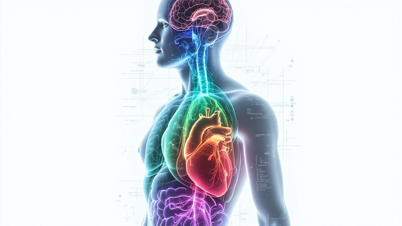
Your organs may be aging at very different speeds — and a simple blood test could soon reveal which ones are falling behind. Researchers at the CeMM Research Center for Molecular Medicine and the Ludwig Boltzmann Institute at the University of Vienna have used artificial intelligence to create tissue clocks that estimate the biological age of individual human organs from histological images.
Published in Nature Medicine, the study analyzed 25,712 tissue images representing approximately 480 million image tiles from 983 individuals across 40 tissue types. The AI models achieved a mean prediction error of just 4.9 years for organ age and revealed that aging does not occur uniformly across the body.
Some organs showed signs of accelerated aging much earlier than expected. Lung, kidney, pancreas, and adrenal gland tissue displayed accelerated aging between ages 20 and 40. The uterus showed a particularly dramatic shift around menopause. Kidney failure was associated with accelerated aging signals across multiple tissues, while diabetes showed pronounced effects specifically in the pancreas.
In a significant conceptual leap, the researchers also built predictors of organ-specific biological age from blood samples alone, by linking blood-based gene expression profiles with the histologically derived tissue age. These blood-based predictors successfully identified aging patterns linked to Alzheimer's disease, Crohn's disease, cystic fibrosis, vasculitis, diabetes, and stroke.
In Alzheimer's disease, the strongest aging signal was detected specifically in the brain, while Crohn's disease showed accelerated aging across the gastrointestinal tract — demonstrating that these tools can capture disease-specific aging signatures.